


Bone Strain Index
SOFTWARE FOR THE AUTOMATIC CALCULATION OF STRESS AND STRAIN OF BONE SEGMENTS
In recent years, the study of bone metabolism has required lot of efforts to take into account fundamental aspects, such as bone microarchitecture, geometry and applied loads, to obtain a correct assessment of bone strength and associated fracture risk.
The tools available today allow a determination of bone density and bone quality, but do not allow to fully analyze the mechanical characteristics of the bone segment under examination.
Bone Strain Index automatically calculates strains and stresses in a bone segment, starting from specific loading conditions for each patient. It is based on finite element calculation algorithms usually used in engineering applications, in the design and test phases of industrial mechanical equipment.
EASY TO USE
Bone Strain Index can be installed on existing DXA densitometers. It receives directly the scan performed with the densitometric examination and analyses the morphological image, the BMD and the body mass of the patient. The time required for the analysis (a few seconds) and the simplicity of use provide to the doctor a useful tool to manage patient data and diagnosis without wasting time.
EASY INTERPRETATION OF RESULTS
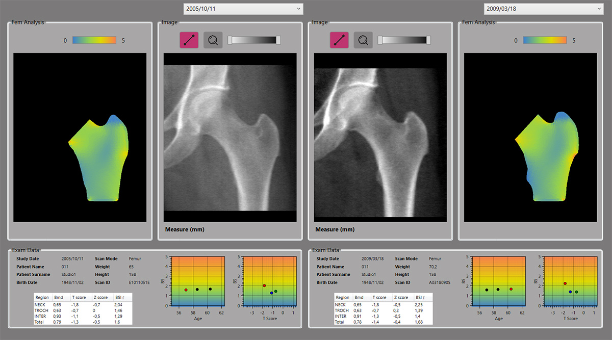
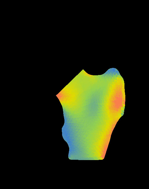
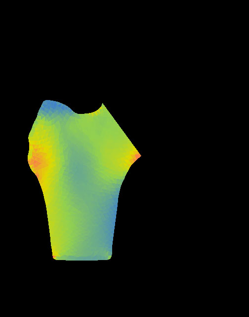
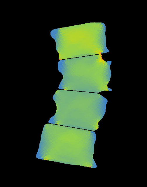
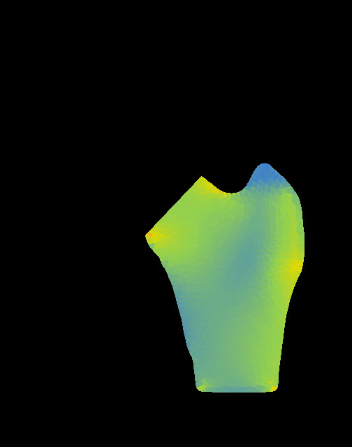
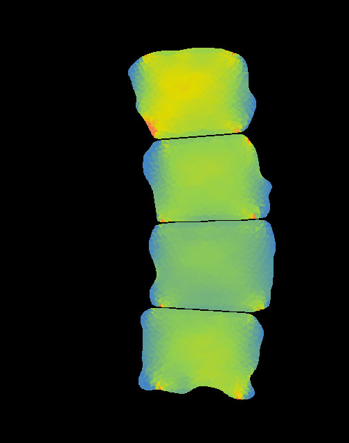
The software provides a fracture risk index called Bone Strain Index and a graphic representation of the deformation distribution in the analyzed bone segment. The colour map follows a ramp from blue (low strain) to green (intermediate strain), yellow, and red (high strain) indicating an increase of the risk factor proportionated to the increase of the strain. In addition, the program provides useful synthetic indicators of the achieved results, such as statistical tables and charts.


VALID DIAGNOSTIC INVESTIGATION TOOL
Resulting data and analysis are a valid tool to support traditional diagnostics and can be applied to both the lumbar and femur regions. Bone Strain Index is complementary to the data available through DXA measurements and may be used in association with the other DXA-derived bone quantity and quality variables for a better risk assessment of patients.
References
Messina C, Piodi LP, Rinaudo L, Buonenna C, Sconfienza LM, Vergani L, Ulivieri FM (2020) Reproducibility of DXA-based bone strain index and the influence of body mass: an in vivo study. Radiol med 125, 313-318 https://doi.org/10.1007/s11547-019-01118-5
SOFTWARE FOR THE AUTOMATIC CALCULATION OF STRESS AND STRAIN OF BONE SEGMENTS
In recent years, the study of bone metabolism has required lot of efforts to take into account fundamental aspects, such as bone microarchitecture, geometry and applied loads, to obtain a correct assessment of bone strength and associated fracture risk.
The tools available today allow a determination of bone density and bone quality, but do not allow to fully analyze the mechanical characteristics of the bone segment under examination.
Bone Strain Index automatically calculates strains and stresses in a bone segment, starting from specific loading conditions for each patient. It is based on finite element calculation algorithms usually used in engineering applications, in the design and test phases of industrial mechanical equipment.
EASY TO USE
Bone Strain Index can be installed on existing DXA densitometers. It receives directly the scan performed with the densitometric examination and analyses the morphological image, the BMD and the body mass of the patient. The time required for the analysis (a few seconds) and the simplicity of use provide to the doctor a useful tool to manage patient data and diagnosis without wasting time.
EASY INTERPRETATION OF RESULTS
The software provides a fracture risk index called Bone Strain Index (BSI) and a graphic representation of the deformation distribution in the analyzed bone segment. The colour map follows a ramp from blue (low strain) to green (intermediate strain), yellow, and red (high strain) indicating an increase of the risk factor proportionated to the increase of the strain. In addition, the program provides useful synthetic indicators of the achieved results, such as statistical tables and charts.
VALID DIAGNOSTIC INVESTIGATION TOOL
Resulting data and analysis are a valid tool to support traditional diagnostics and can be applied to both the lumbar and femur regions. BSI is complementary to the data available through DXA measurements and may be used in association with the other DXA-derived bone quantity and quality variables for a better risk assessment of patients.
References
Tabacco G, Naciu AM, Messina C, Sanson G, Rinaudo L, Cesareo R, Falcone S, Manfrini S, Napoli N, Bilezikian JP, Ulivieri FM, Palermo A. DXA-Based Bone Strain Index: A New Tool to Evaluate Bone Quality in Primary Hyperparathyroidism. J Clin Endocrinol Metab. 2021 May 8:dgab317. doi: 10.1210/clinem/dgab317
Messina C, Piodi LP, Rinaudo L, Buonenna C, Sconfienza LM, Vergani L, Ulivieri FM (2020) Reproducibility of DXA-based bone strain index and the influence of body mass: an in vivo study. Radiol med 125, 313-318 https://doi.org/10.1007/s11547-019-01118-5