DICOM Viewers
ITA WEB – A WEB-BASED VIEWER FOR ITA PACS
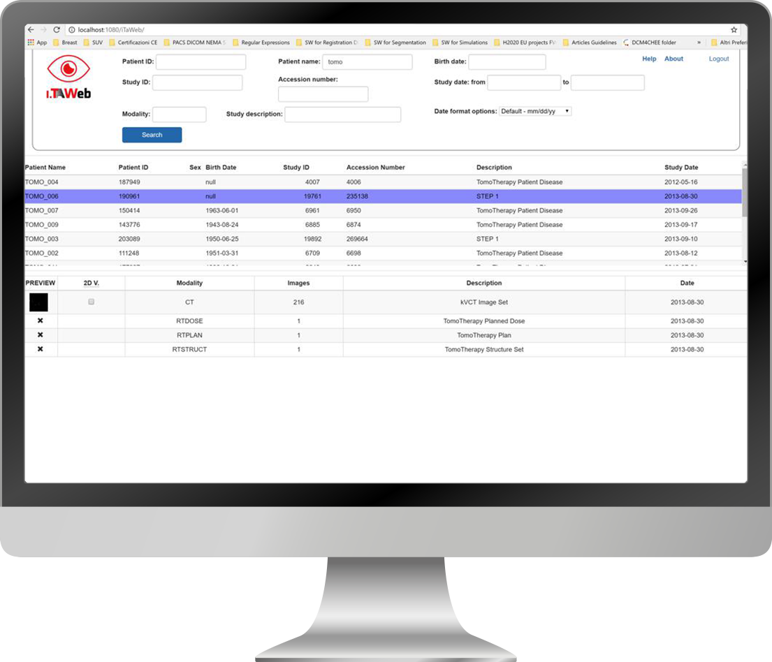
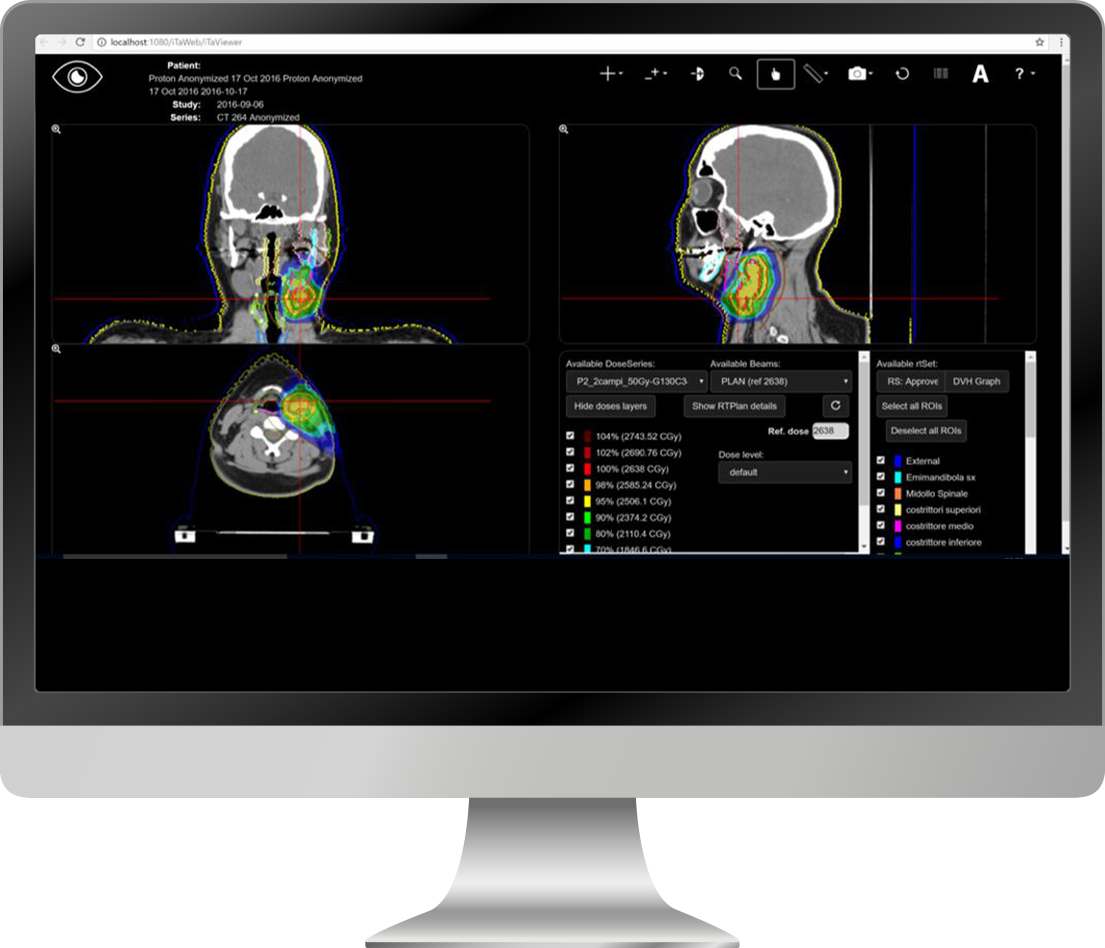
iTA Web is a web-based viewer, which uses HTML5 and JavaScript for processing and displaying of 3D images. Images are DICOM P10, they are transferred via HTTP(s) protocol, processed and displayed by browser. Supported browser: Google Chrome, Mozilla Firefox, Opera, Apple Safari, Microsoft IE10+, Microsoft Edge. The interface is also optimized for mobile devices (tablets).
iTA Web allows to:
- query iTA PACS DB to select the study;
- display CT/PET/MR DICOM series in MPR modality (coronal, sagittal e transverse), RTSTRUCT and RTDOSE;
- zoom, pan, adjust brightness/contrast image value;
- display DVHs;
- select images, add notes and graphic elements (arrow, point, line) to image and generate reports.
ITA VIEW – DICOM VIEWER FOR IPAD
iTA VIEW is a viewer for DICOM images and DICOM RT objects designed for radiotherapy departments.
iTA VIEW allows you to connect to any DICOM node, and to import Studies into the local database.
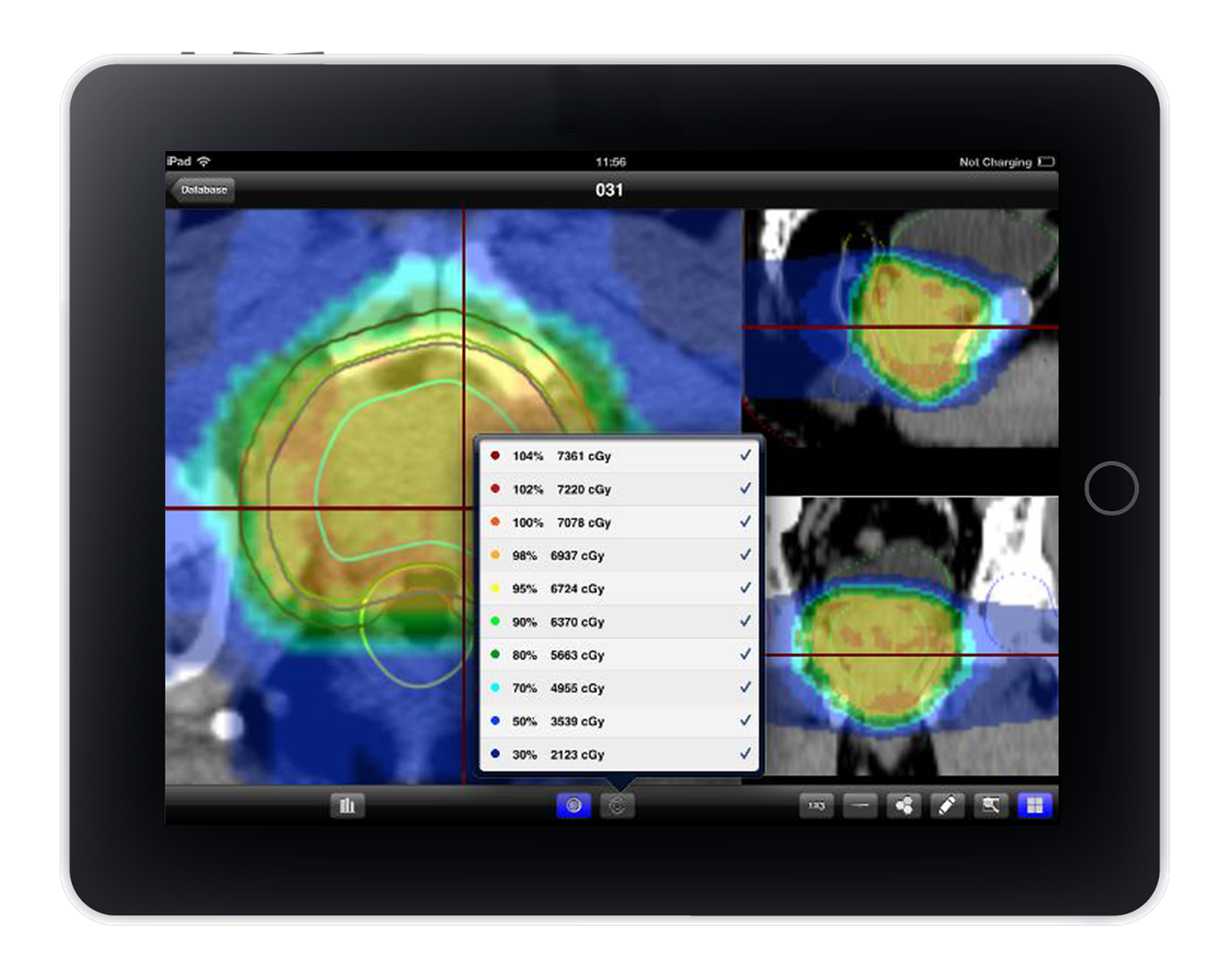
With iTA VIEW you can display CT, MR, PT and 3DUS images and DICOM RT objects, such as RT Images, set of structures, dose distributions and DVHs. The original images (axial view) and the reconstructed planes (sagittal and coronal views) are visualized. The resolution of the reconstructed views is chosen by the user. The Regions of Interest (ROI) contours and dose distributions are displayed on the three views.
iTA VIEW calculates the dose-volume histograms (DVH) and displays the results for the selected ROIs. The DVHs are shown both in absolute and relative dose; selecting a point on the DVH, its dose-volume values are displayed.
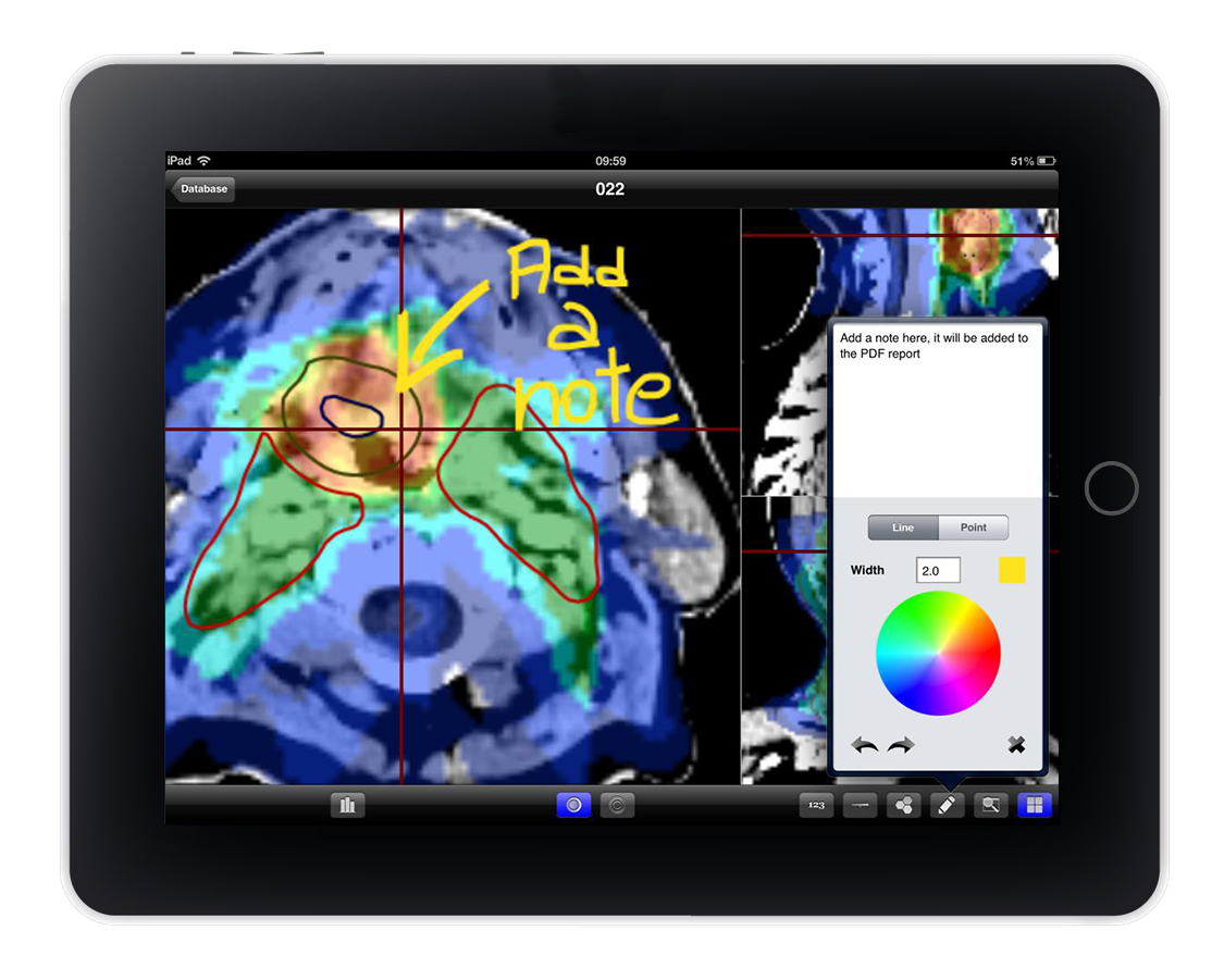
iTA VIEW provides tools for drawing and writing that can be used to add comments, lines, arrows and points to the axial views. The pictures with comments and notes are added to the PDF report that is created by the application and which also contains basic information about the patient and the treatment plan and DVHs. Also pictures (patient, positioning system, etc.) and information encoded in barcodes can be added to the PDF report.
iTA VIEW CANNOT be used as a medical device for primary diagnosis.
Functionalities:
- Query/retrieve from all DICOM servers (C-Get, C-Move).
- Import in local database of studies via iTunes File Sharing.
- RSearch and show Studies in local database (with the free version only two Studies at a time can be stored in the local database).
- Visualization of DICOM images (CT, PT, MR, 3DUS), DICOM RT Images and DICOM RT Objects (ROIs, POIs and DOSEs).
- Traditional 2D visualization: axial, sagittal and coronal views.
- Visualization of ROIs from DICOM RT Structures.
- Zoom and Pan.
- Contrast and intensity adjustment.
- 2D dose visualization from DICOM RT Dose.
- Custom isodoses visualization.
- DVH calculation and visualization (available in the full version).
- Tools for freehand drawing and writing notes on images.
- Take and add pictures (patient, positioning system, etc.) to the PDF report.
- Scan a barcode (or use a photo) and add the encoded information to the PDF report.
- Send reports by email, skype, etc. (in PDF format) or to a DICOM node (DICOM – DOC modality) with selected images, DVH(s), information about the patient and the treatment plan, pictures and barcode information (available in the full version).
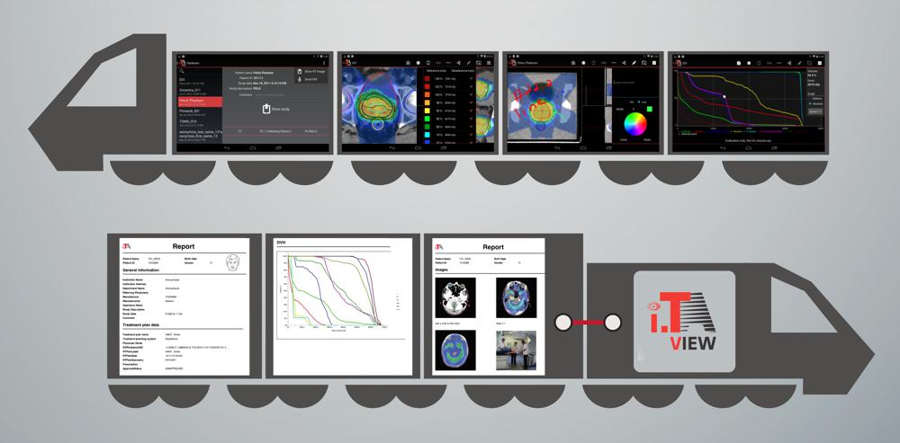

iTA VIEW è un visualizzatore di oggetti DICOM e DICOM RT sviluppato per iPad e dedicato alla radioterapia. iTA VIEW permette di connettersi a qualsiasi nodo DICOM per l’importazione degli studi.
Con iTA VIEW è possibile sia visualizzare immagini CT, MR, PET e 3DUS sia oggetti DICOM RT, quali RT Image, set di strutture, distribuzioni di dosi e DVH. Vengono visualizzate sia le immagini originali (vista assiale) sia i piani ricostruiti (vista sagittale e vista coronale). La risoluzione delle viste ricostruite viene scelta dall’utente. I contorni delle Regioni di Interesse (ROI) e le distribuzioni di dose vengono visualizzati sulle tre viste.
iTA VIEW calcola gli istogrammi Dose-Volume (DVH) e ne visualizza il risultato per le ROI selezionate. È possibile selezionare diversi punti sul DVH e visualizzarne i valori dose-volume. iTA VIEW è dotato di strumenti di disegno e scrittura che possono essere utilizzati per aggiungere commenti, linee, frecce e punti alle viste assiali. Le immagini con note e commenti vengono aggiunte al report PDF che viene creato dall’applicazione e che contiene anche le informazioni principali relative al paziente e al piano di trattamento e i DVH. È possibile aggiungere al report PDF anche foto e informazioni contenute in codici a barre.
iTA VIEW NON può essere utilizzato come Dispositivo Medico per diagnosi primaria.
Functionalities:
- Query/retrieve from all DICOM servers (C-Get, C-Move).
- Import in local database of studies via iTunes File Sharing.
- RSearch and show Studies in local database (with the free version only two Studies at a time can be stored in the local database).
- Visualization of DICOM images (CT, PT, MR, 3DUS), DICOM RT Images and DICOM RT Objects (ROIs, POIs and DOSEs).
- Traditional 2D visualization: axial, sagittal and coronal views.
- Visualization of ROIs from DICOM RT Structures.
- Zoom and Pan.
- Contrast and intensity adjustment.
- 2D dose visualization from DICOM RT Dose.
- Custom isodoses visualization.
- DVH calculation and visualization (available in the full version).
- Tools for freehand drawing and writing notes on images.
- Take and add pictures (patient, positioning system, etc.) to the PDF report.
- Scan a barcode (or use a photo) and add the encoded information to the PDF report.
- Send reports by email, skype, etc. (in PDF format) or to a DICOM node (DICOM – DOC modality) with selected images, DVH(s), information about the patient and the treatment plan, pictures and barcode information (available in the full version).


